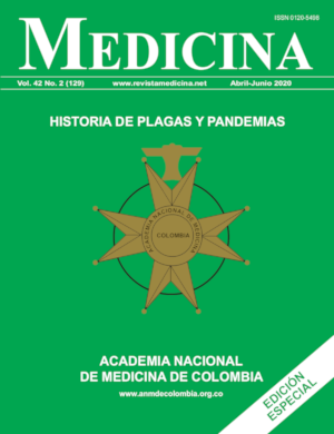
La tercera pandemia de peste de 1855
DOI:
https://doi.org/10.56050/01205498.1518Palabras clave:
tercera pandemia, Yersinia Pestis, respuesta inmunológica, zoonosis, Rattus rattus, Rattus norvegicus, Xenopsylla cheopis, Ctenocephalides felis, Pulex irritansResumen
El presente escrito surge durante la cuarentena, aceptada por el pueblo colombiano desde el mes de marzo y siguientes del año común 2020 por disposición gubernamental, para disminuir el riesgo de contagio y de muerte durante la Pandemia del “Coronavirus”, SARS-CoV-2, Covid19.
Todas las epidemias, plagas y pandemias conocidas sobre la faz de la tierra, han formado parte de la evolución y la naturaleza humana. Hoy, en pleno siglo XXI, después de haber viajado al espacio, haber conquistado la Luna, de tener medios de comunicación instantáneos, “casi telepáticos”, una estructura proteica virtualmente invisible, microscópica, “inerte”, nos mantiene confinados en el mismo tiempo, a más de la mitad de los 7.800 millones de Seres Humanos que cohabitamos en el Planeta Azul. Nada ni nadie nos había exhortado a tantas personas juntas a detenernos durante horas, días, quizá semanas o aún meses, a pensar en lo insignificantes que somos ante el implacable poder de la Naturaleza.
Biografía del autor/a
María Claudia Ortega López, Hospital Infantil Universitario de San José
Referencias bibliográficas
2. Morelli G, Song Y, Mazzoni CJ, Eppinger M, Roumagnac P, Wagner DM, et al. Yersinia pestis genome sequencing identifies patterns of global phylogenetic diversity. Nat Genet. 2010; 42(12):1140– 3. Epub 2010/11/03. 10.1038/ng.705 [pii] 10.1038/ng.705.
3. Wagner DM, Klunk J, Harbeck M, Devault A, Waglechner N, Sahl JW, et al. Yersinia pestis and the Plague of Justinian 541–543 AD: a genomic analysis. The Lancet Infectious diseases. 2014; 14(4):319– 26. 10.1016/S1473- 3099(13)70323-2.
4. Perry RD, Fetherston JD. Yersinia pestis-etiologic agent of plague. Clin Microbiol Rev. 1997; 10(1):35–66. Epub 1997/01/01.
5. Raoult D, Mouffok N, Bitam I, Piarroux R, Drancourt M. 2013. Plague: history and contemporary analysis. J. Infect. 66, 18–26. (10.1016/j.jinf.2012.09.010).
6. Whittles LK, Didelot X. Epidemiological analysis of the Eyam plague outbreak of 1665– 1666. 2016. Proc. R. Soc. B 283, 20160618 (10.1098/rspb.2016.0618).
7. Xavier Didelot, Lilith K. Whittles, and Ian Hall. Model-based analysis of an outbreak of bubonic plague in Cairo in 1801. J R Soc Interface. 2017 Jun; 14(131): 20170160. PMCID: PMC5493801 Published online 2017 Jun 21. Doi: 10.1098/rsif.2017.0160 PMID: 28637916.
8. Rotz LD, Khan AS, Lillibridge SR, Ostroff SM, Hughes JM. Public health assessment of potential biological terrorism agents. Emerg Infect Dis. 2002; 8(2):225–30. Epub 2002/03/19. 10.3201/eid0802.010164.
9. Slack P. Plague: a very short introduction. Oxford, UK: Oxford University Press 2012. ISBN: 9780199589548.
10. Devaux CA. Small oversights that led to the Great Plague of Marseille (1720–1723): lessons from the past. Infect. Genet. Evol. 14, 169–185. (10.1016/j.meegid.2012.11.016). 2013.
11. Cummins N, Kelly M, Ó Gráda C. Living standards and plague in London, 1560–1665. Econ. Hist. Rev. 69, 3–34. (10.1111/ehr.12098). 2016.
12. Pryor EG. The great plague of Hong Kong. J. Hong Kong Branch R. Asiat. Soc. 1975; 15, 61–70.
13. Butler T. Plague history: Yersin’s discovery of the causative bacterium in 1894 enabled, in the subsequent century, scientific progress in understanding the disease and the development of treatments and vaccines. Clin. Microbiol. Infect. 20, 202–209. (10.1111/1469-0691.12540) 2014.
14. WHO. Plague-Madagascar, Disease outbreak news. 2017, http://www.who.int/csr/don/09-january-2017-plague-mdg/en/.
15. Dos Santos Grácio AJ, Grácio MA. Plague: A Millenary Infectious Disease Reemerging in the XXI Century. Biomed Res Int. 2017(33):1-8. Published online 2017 Aug 20. doi: 10.1155/2017/5696542.
16. Bertherat E. Plague around the world, 2010–2015. Wkly Epidemiol Rec. 2016; 91:89–104.
17. Respicio-Kingry LB, Yockey BM, Acayo S, Kaggwa J, Apangu T, Kugeler KJ, et al. Two distinct Yersinia pestis populations causing plague among humans in the West Nile region of Uganda. PLoS Negl Trop Dis. 2016; 10:e0004360.
18. Shi L, Yang G, Zhang Z, Xia L, Liang Y, Tan H, et al. Reemergence of human plague in Yunnan, China in 2016. PLoS ONE. 2018; 13:e0198067.
19. Abedi AA, Shako J-C, Gaudart J, Sudre B, Ilunga BK, Sha- mamba SKB, et al. Ecologic features of plague outbreak areas, Democratic Republic of the Congo, 2004– 2014. Emerg Infect Dis. 2018; 24:210-20.
20. Andrianaivoarimanana V, Piola P, Wagner DM, Rakotomanana F, Maheriniaina V, Andrianalimanana S, et al. Trends of human plague, Madagascar, 1998–2016. Emerg Infect Dis. 2019; 25:220-8.
21. McNally A, Thomson NR, Reuter S, Wren BW. ‘Add, stir and reduce’: Yersinia spp. as model bacteria for pathogen evolution. Nat Rev Micro. 2016; 14:177-90.
22. Christian E. Demeure, Olivier Dussurget Guillem Mas Fiol, Anne-Sophie Le Guern, Cyril Savin, Javier PizarroCerdá Yersinia pestis and plague: an updated view on evolution, virulence determinants, immune subversion, vaccination, and diagnostics. Genes and Immunity 2019; 20:357-370 https://doi.org/10.1038/s41435-019-0065-0.
23. Spinner JL, Winfree S, Starr T, Shannon JG, Nair V, Steele- Mortimer O, et al. Yersinia pestis survival and replication within human neutrophil phagosomes and uptake of infected neutrophils by macrophages. J Leukoc Biol. 2014; 95:389–98.
24. Hinnebusch BJ, Jarrett CO, Bland DM. ‘Fleaing’ the plague: adaptations of Yersinia pestisto its insect vector that lead to transmission. Annu Rev Microbiol. 2017; 71:215– 32.
25. Shannon JG, Hasenkrug AM, Dorward DW, Nair V, Carmody AB, Hinnebusch BJ. Yersinia pestis subverts the dermal neutrophil response in a mouse model of bubonic plague. MBio. 2013; 4:e00170-13-e00170-13.
26. Pujol C, Klein KA, Romanov GA, Palmer LE, Cirota C, Zhao Z, et al. Yersinia pestis can reside in autophagosomes and avoid xenophagy in murine macrophages by preventing vacuole acidification. Infect Immun. 2009; 77:2251–61.
27. Connor MG, Pulsifer AR, Price CT, Abu Kwaik Y, Lawrenz MB. Yersinia pestis requires host Rab1b for survival in macrophages. PLoS Pathog. 2015; 11:e1005241.
28. Connor MG, Pulsifer AR, Chung D, Rouchka EC, Ceresa BK, Lawrenz MB. Yersinia pestis targets the host endosome recycling pathway during the biogenesis of the Yersinia-containing vacuole to avoid killing by macrophages. MBio. 2018;9:e01800-17
29. Merritt PM, Nero T, Bohman L, Felek S, Krukonis ES, Marketon MM. Yersinia pestis targets neutrophils via complement receptor 3. Cell Microbiol. 2014; 17:666-87.
30. Derbise A, Pierre F, Merchez M, Pradel E, Laouami S, Ricard I, et al. Inheritance of the lysozyme inhibitor Ivy was an important evolutionary step by Yersinia pestis to avoid the host innate immune response. J Infect Dis. 2013; 207:1535-43.
31. Reboul A, Lemaître N, Titecat M, Merchez M, Deloison G, Ricard I, et al. Yersinia pestis requires the 2-component regulatory system OmpR-EnvZ to resist innate immunity during the early and late stages of plague. J Infect Dis. 2014; 210:1367-75.
32. Arifuzzaman M, Ang WXG, Choi HW, Nilles ML, St John AL, Abraham SN. Necroptosis of infiltrated macrophages drives Yersinia pestis dispersal within buboes. JCI Insight. 2018; 3:35.
33. St. JohnAL,AngWXG, Huang M-N,Kunder CA, ChanEW, Gunn MD, et al. S1P-dependent trafficking of intracellular Yersiniapestisthroughlymphnodesestablishesbuboesand systemic infection. Immunity. 2014; 41:440-50.
34. Littman DR, Rudensky AY. Th17 and regulatory T cells in mediating and restraining inflammation. Cell. 2010; 140:845-58.
35. Bi Y, Zhou J, Yang H, Wang X, Zhang X, Wang Q, et al. IL17A produced by neutrophils protects against pneumonic plague through orchestrating IFN-γ-activated macrophage programming. J Immunol. 2014; 192:704-13.
36. Lin JS, Kummer LW, Szaba FM, Smiley ST. IL-17 contributes to cell-mediated defense against pulmonary Yersinia pestis infection. J Immunol. 2011; 186:1675-84.
37. Comer JE, Sturdevant DE, Carmody AB, Virtaneva K, Gardner D, Long D, Rosenke R, Porcella SF, Hinnebusch BJ. 2010. Transcriptomic and innate immune responses to Yersinia pestis in the lymph node during bubonic plague. Infect. Immun. 78:5086-5098.
38. Sebbane F, Gardner D, Long D, Gowen BB, Hinnebusch BJ. 2005. Kinetics of disease progression and host response in a rat model of bubonic plague. Am. J. Pathol. 166:1427-1439.
39. Pechous RD, Sivaraman V, Price PA, Stasulli NM, Goldman WE. Early host cell targets of Yersinia pestis during primary pneumonic plague. PLoS Pathog. 2013; 9:e1003679.
40. Peters KN, Dhariwala MO, Hughes Hanks JM, Brown CR, Anderson DM. Early apoptosis of macrophages modulated by injection of Yersinia pestis YopK promotes progression of primary pneumonic plague. PLoS Pathog. 2013; 9:e1003324.
41. Stasulli NM, Eichelberger KR, Price PA, Pechous RD, Montgomery SA, Parker JS, et al. Spatially distinct neutrophil responses within the inflammatory lesions of pneumonic plague. MBio. 2015; 6:e01530-15.
42. WHO. How to safely collect sputum samples from patients suspected to be infected with pneumonic plague. 2016. Technical Guidance. www.who.int/csr/disease/plague/en.
43. Tourdjman M, Ibraheem M, Brett M, DeBess E, Progulske B, Ettestad P, et al. Misidentification of Yersinia pestis by automated systems, resulting in delayed diagnoses of human plague infections Oregon and New Mexico, 2010– 2011. Clin Infect Dis. 2012; 55:e58–e60.
44. Morelli G, et al. Yersinia pestis genome sequencing identifies patterns of global phylogenetic diversity. Nat Genet 2010; 42:1140–1143.
45. Achtman M, et al. Yersinia pestis, the cause of plague, is a recently emerged clone of Yersinia pseudotuberculosis. Proc Natl Acad Sci USA. 1999; 96:14043-14048.
46. Lei Xu, Leif C. Stige, Herwig Leirs, Simon Neerinckx, Kenneth L. Gage, Ruifu Yang, Qiyong Liu, Barbara Bramanti, Katharine R. Dean, Hui Tang, Zhe Sun, Nils Chr. Stenseth, and Zhibin Zhang. Historical and genomic data reveal the influencing factors on global transmission velocity of plague during the Third Pandemic. Proc Natl Acad Sci USA. 2019 Jun 11; 116(24): 11833-11838.
47. BramantiB,DeanKR,Walløe L,Chr.StensethN. 2019The Third Plague Pandemic in Europe. Proc. R. Soc. B 286: 20182429. http://dx.doi.org/10.1098/rspb.2018.2429.
48. Pollitzer R. Division of Epidemiology, World Health Organization. Plague Studies. A Summary of the History and a Survey of the Present Distribution of the Disease. Bull. Org. mond. Santé. Bull. World Hlth Org. 1951; 4:475-533.
49. Scheube B. The diseases of warm countries: A handbook for medical men. London, UK: Bale & Danielsson. 1908.
50. Faccini-Martínez A, Sotomayor HA. Reseña histórica de la peste en Suramérica: una enfermedad poco conocida en Colombia. Biomédica. 2013; 33(1): 8-27. https://doi.org/10.7705/biomedica.v33i1.814.
Cómo citar
Descargas
Publicado
Número
Sección
Licencia
Derechos de autor 2020 Medicina

Esta obra está bajo una licencia internacional Creative Commons Atribución-NoComercial-SinDerivadas 4.0.
Copyright
ANM de Colombia
Los autores deben declarar revisión, validación y aprobación para publicación del manuscrito, además de la cesión de los derechos patrimoniales de publicación, mediante un documento que debe ser enviado antes de la aparición del escrito. Puede solicitar el formato a través del correo revistamedicina@anmdecolombia.org.co o descargarlo directamente Documento Garantías y cesión de derechos.docx
Copyright
ANM de Colombia
Authors must state that they reviewed, validated and approved the manuscript's publication. Moreover, they must sign a model release that should be sent.


